Venous Valves on Ultrasound

Blogs
Blogs
Venous Valves on Ultrasound
")
Valves are one of the key components of normal venous function. There are valves throughout the veins in the body. They control the direction of blood flow and are extremely important in the movement of blood out of the legs back to the heart. Valves consist of two flaps (cusps or leaflets) with edges that meet. They allow blood to flow in only one direction - towards the heart. One of the best tools to look at how the valves are function is ultrasound (US). US allows us to look at the structure of the valve and how it is functioning. US allows us to see how blood is moving by applying color to moving blood.
Click to see the valves in action.
Below is series of images demonstrating normal valve function.
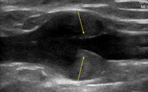
This US image shows the valve leaflets (yellow arrows) open. When the valve is open blood is flowing from the leg back to the heart.
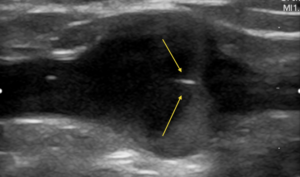
The next US images shows the leaflets to be closed. The yellow arrows show the 2 leaflets meeting together. This prevents the blood the just moved toward the heart from flowing back down the leg.
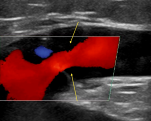
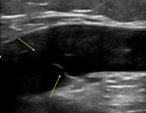
Valves may stop working due to a variety of mechanisms (often hereditary or after a blood clot). The image below shows the 2 leaflets (yellow arrows) not meeting together. This allows blood to flow the wrong direction in the vein.
Using color, we can see blood leaking through the valve in the wrong direction (red). The yellow arrows show the jet of blood (red color) as it passes through the 2 valve leaflets that are not closing correctly.